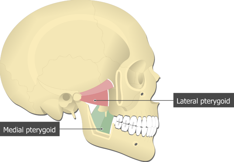
Each pterygoid process projects inferiorly from the junction of the body and greater wing of the sphenoid bone and bifurcates into a medial pterygoid plate and a lateral pterygoid plate. The superficial head is attached to the maxillary tuberosity and the pyramidal process of the palatine bone.
Medial Pterygoid Learn Muscles
Using the medial and lateral pterygoid muscles as references identify the buccal branch of the mandibular nerve CN V3 and accompanying buccal artery.

. The medial pterygoid lies on the medial side of the mandible as a mirror image of the masseter. This origin is medial to the insertion so can contribute to rotating the mandible causing some side to side movement. It also assists the lateral pterygoid muscle with side to side mandibular motion to help with the grinding of food.
It depresses and protrudes the mandible. The smaller superficial head has its origin mainly from the maxillary tuberosity with some fibers arising from the pyramidal process of the palatine bone. Lateral Pterygoid The lateral pterygoid muscle is the primary muscle of the inferior temporal fossa.
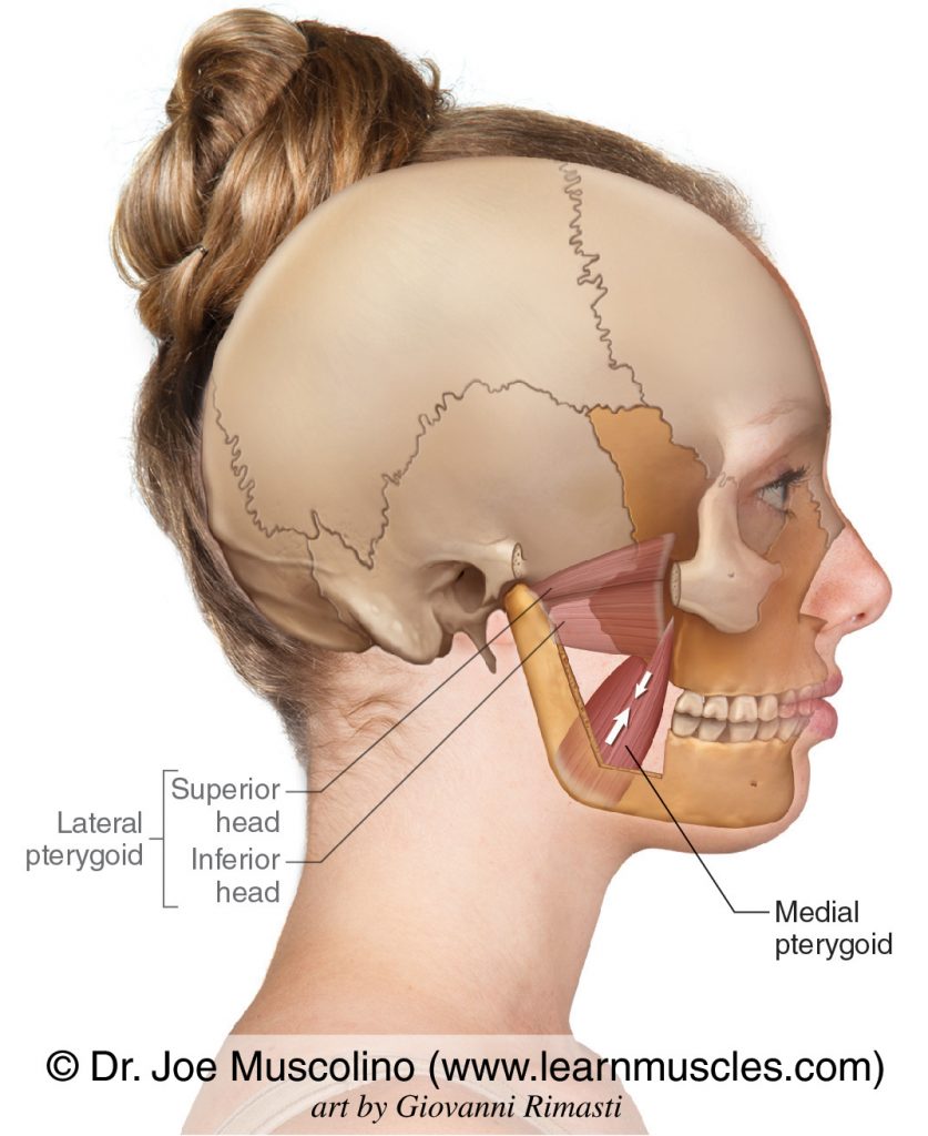
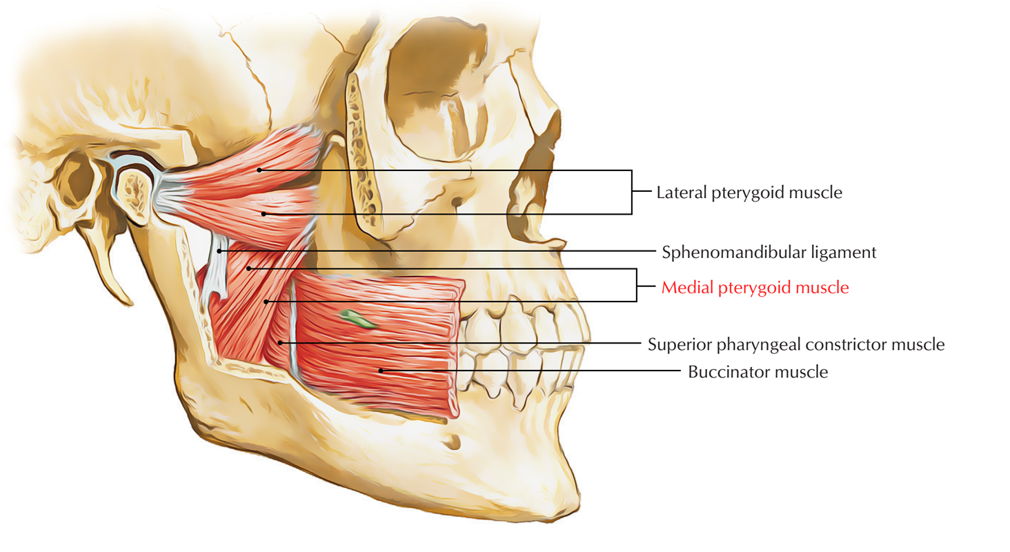
Branches of the mandibular division of the trigeminal nerve CN V3 Blood supply. The medial pterygoid muscle attaches to the angle of the mandible and to the lateral pterygoid plate to form a sling with the masseter muscle that suspends the mandible Figure 6-19. The nerve and artery usually pass between the two heads of the lateral pterygoid muscle.
It has two heads of origin. The medial pterygoid muscle is a thick and square shaped muscle. The lateral pterygoids activate at an angle to one another and push the mandible forward.
It can assist in protrusion of the mandible. The lateral pterygoid muscle or external pterygoid muscle is a muscle of mastication. The pterygoids are often weak and inactive due to the underdevelopment of the maxilla which causes occlusion to establish too posteriorlyThe pterygoids pro.
Using the medial and lateral pterygoid muscles as references identify the buccal branch of the mandibular nerve CN V3 and accompanying buccal artery. The nerve and artery usually pass between the two heads of the lateral pterygoid muscle. The medial pterygoid muscle functions to assist with elevation and protrusion of the mandible.
Although having different origins both heads insert on the inner. Like the medial pterygoid and the masseter it has two heads. The bulk of the muscle arises as a deep head from just above the medial surface of the lateral pterygoid plate.
It lies superior to the medial pterygoid muscle. The medial pterygoid muscle has a superficial and a deep head which arise from different areas of the jaw. The deep head is the major component and is attached to the medial aspect of the lateral pterygoid plate of the sphenoid bone.
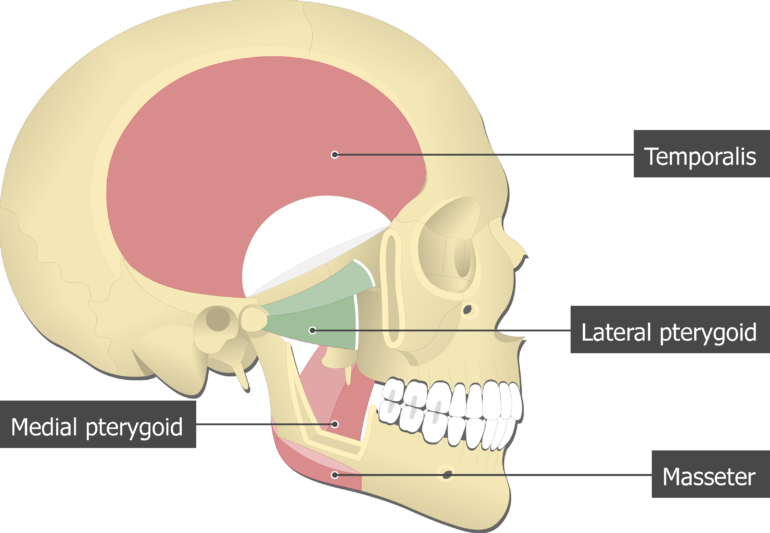
At the inferior tip of the medial pterygoid plate is the small hook-shaped process the pterygoid hamulus. These two nerves pass between the medial and lateral pterygoid muscles. Medial and Lateral Pterygoids are both well hidden by the lower jaw bone.
The medial pterygoid can be very hyperactive in jaw closing dystonia as a result of the so-called whack-a-mole phenomenon after repeated botulinum toxin injections into the masseter and temporalis muscles. Jaw opening jaw deviation and jaw protrusion types of oromamdibular dystonia are caused by involuntary contraction of the lateral pterygoid muscles. The medial pterygoid muscle consists of two heads.
The primary action is to elevate the mandible and laterally deviate it to the opposite side. The deep part originates on the medial surface of the lateral pterygoid plate and inserts in the same area of the mandible. The medial pterygoids temporalis and masseter muscles elevate the mandible to close the jaw and the lateral pterygoids help open it other muscles involved in jaw opening are the digastric and geniohyoid muscles.
Lateral pterygoid medial pterygoid. Pterygoid branches of the maxillary artery. The lateral pterygoid muscle via its lower head separates the two distinct heads.
The superior head originates on the sphenoid bone facing the infratemporal fossa. Protrusion elevation medial movement of mandible. Medial pterygoid is a thick quadrilateral muscle that connects the mandible with maxilla sphenoid and palatine bones.
It is supplied by pterygoid branches of the maxillary artery and the lateral pterygoid nerve from the mandibular nerve CN V 3. Both heads originate anterior to the insertions so like the superficial head of the masseter it is. These two nerves pass between the medial and lateral pterygoid muscles.
Click to see full answer. Using the medial and lateral pterygoid muscles as references identify the buccal branch of the mandibular nerve CN V3 and accompanying buccal artery. When each muscle works independently they can move the.
Lateral Pterygoid this muscle lives directly behind your upper back molar using the same-sided hand ie right hand for right pterygoid with your palm against your cheek place your pinky inside your cheek on the row of upper teeth your jaw can rest closed once. It belongs to the group of masticatory muscles along with the lateral pterygoid masseter and temporal muscles. It arises from the pterygoid fossa of the sphenoid bone and the lateral lamina of the pterygoid process and ascends to the inner surface of the mandibular angle and pterygoid tuberosity.
These two nerves pass between the medial and lateral pterygoid muscles. The nerve and artery usually pass between the two heads of the lateral pterygoid muscle. The medial pterygoid muscle causes pain in the temporomandibular joint and the ear which increases when you bite down on something.
Tightness in this muscle can make it difficult to open the mouth wide. The smaller superficial head originates from the maxillary tuberosity and the pyramidal process of the palatine bone. The lateral pterygoid is the big exception in this muscle group in that its fibers run horizontally rather than vertically and one of the two heads of the muscle does not insert into bone but rather inserts into the articular cartilage of the TMJ.
Lateral pterygoid TrPs Refer often severe pain into the maxillary sinus deeply into the TM joint produce dysfunctions of the joint. Medial pterygoid muscle consists of two heads. It has two heads.
Slightly reduced range of opening deviations of mandible with opening altered occlusion my teeth dont fit together right tinnitus are common complaints associated with TrPs in this muscle. Protrusion depression medial movement of mandible.
Medial Pterygoid Muscle Earth S Lab
Lateral Pterygoid Muscle Attachments Actions Innervation

Lateral Pterygoid Muscle Wikipedia
Lateral Pterygoid Muscle Wikipedia
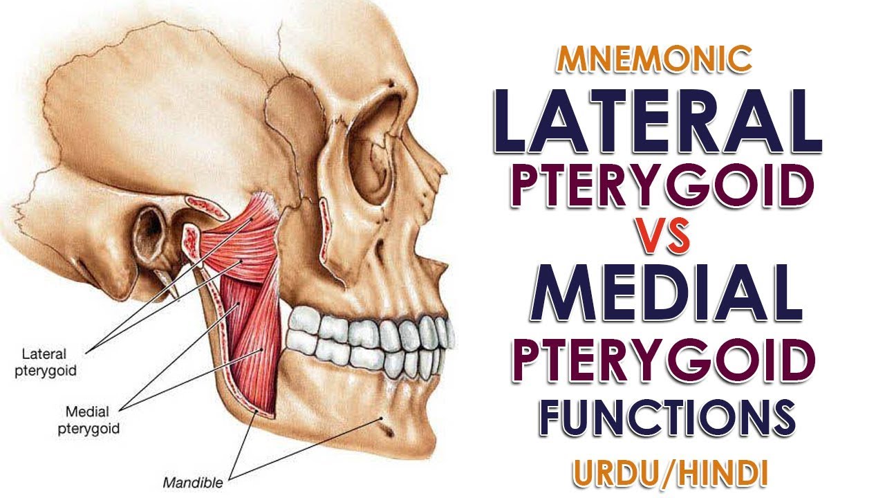
Mnemonic Lateral Pterygoid Vs Medial Pterygoid Function Urdu Hindi Youtube
Medial Pterygoid Muscle Attachments Actions Innervation
![]()
Medial And Lateral Pterygoid Muscle Anatomy And Function Kenhub
![]()
Medial And Lateral Pterygoid Muscle Anatomy And Function Kenhub
0 comments
Post a Comment